Смотреть видео: Как выглядит клетчатка под микроскопом
Как выглядит клетчатка под микроскопом - найдено 60 фото
Найдено фото: 60
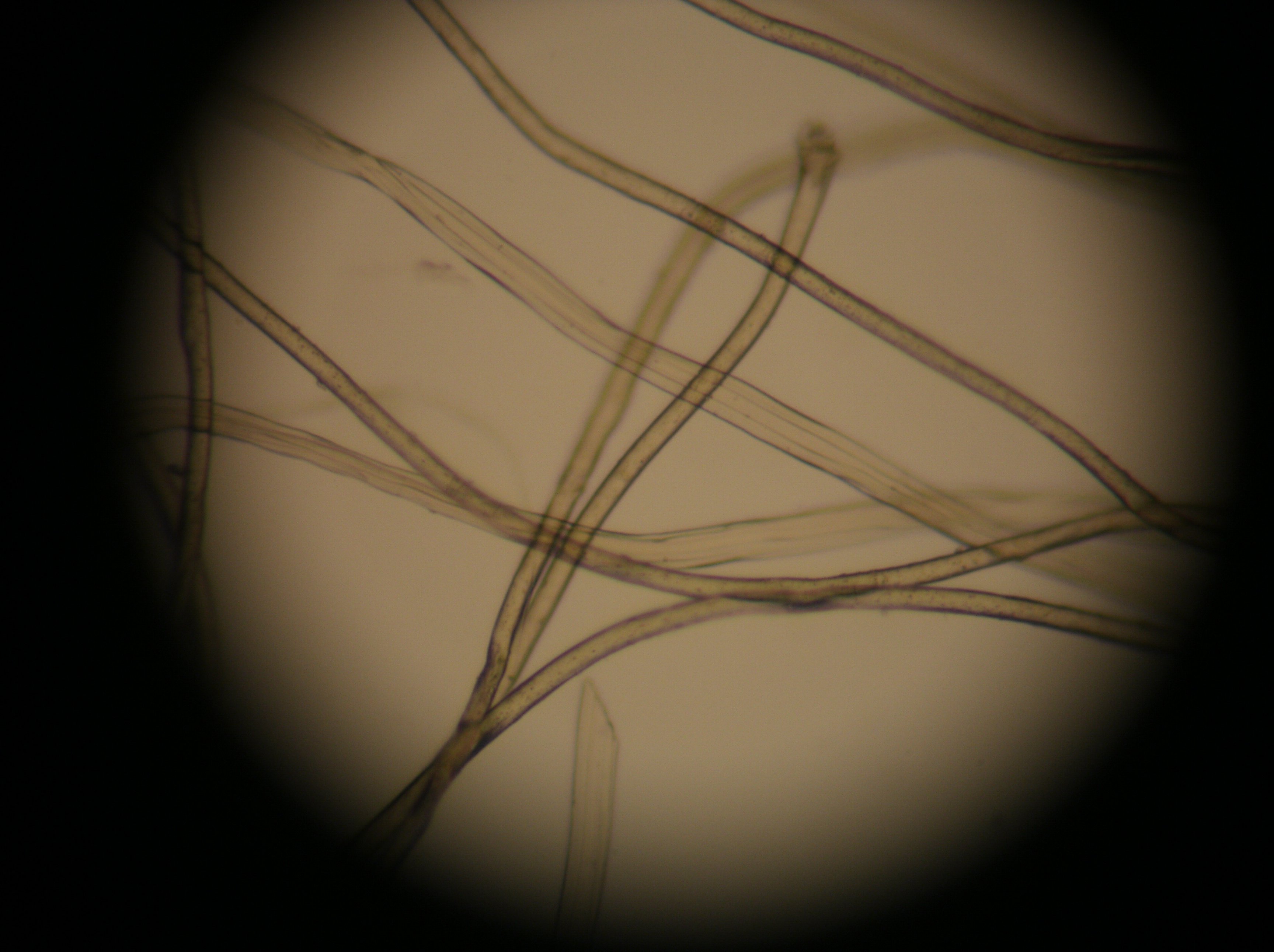
Cellulose fibres (paper towel), SEM - Stock Image - C032/5018 - Science Photo LiУченые узнали больше о беспорядочных связях липидов и белков
School of Engineering Vanderbilt UniversityМикроскоп Микромед Р-1 LED купить от официального дилера с гарантиейЕда под микроскопом - Статьи на сайте Четыре глазаFood under the microscope: scanning electron micrographs of foodstuffs Scanning Клетчатка неперевариваемая - CoffeePapa.ruNever Mind Lasagnas: All Food Is Pretty Gross Up Close... Electron microscope, M
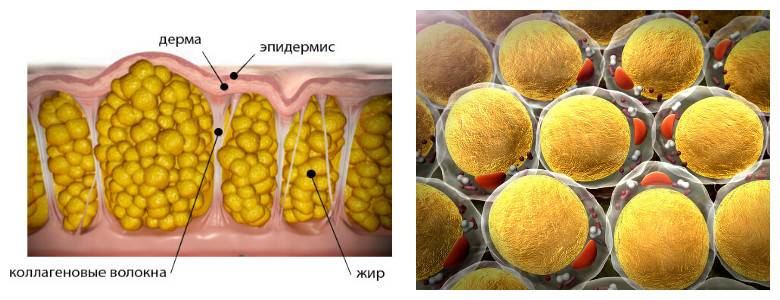
Gallery Image Patterns in nature, Futuristic art, Electron microscopeclassification " Year 7 Science - Mr WrightFreeze fraction of a Coffee bean showing the content of the cells. Scanning ElecPoussière domestique Scanning electron microscope images, Electron microscope imЖиры в организме человека. Виды жира в теле: висцеральный жир, подкожный, бурый,Pin on Healthy Weight Loss
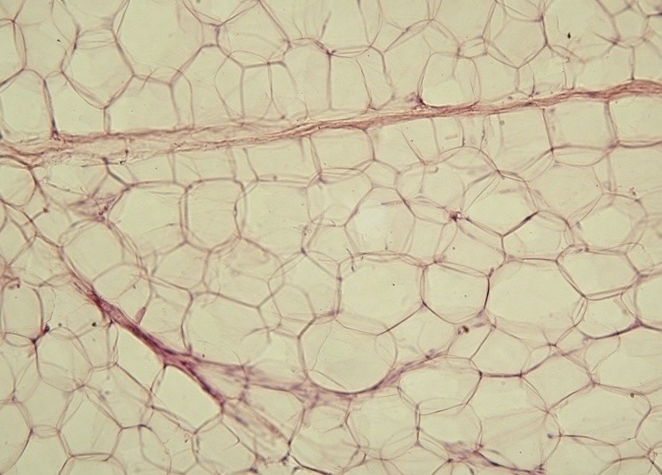
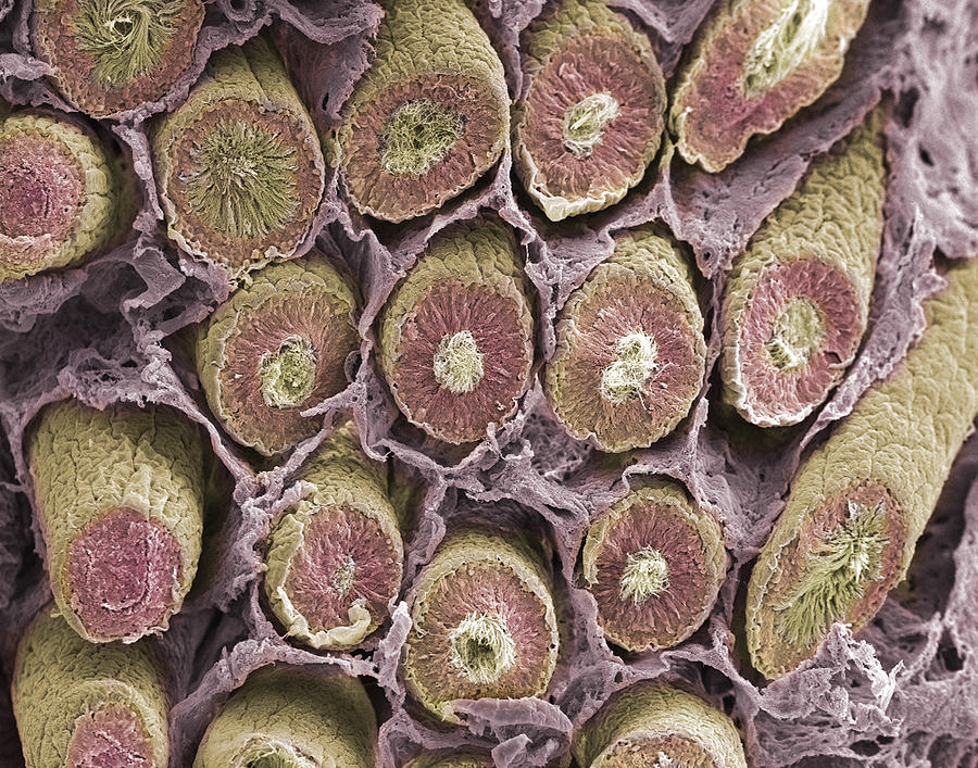
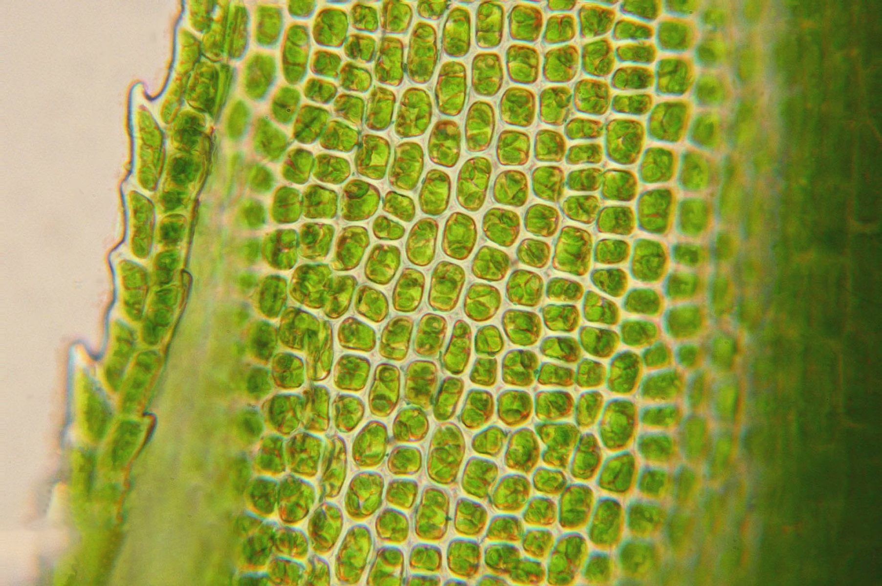
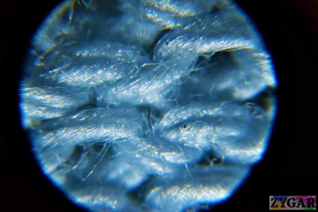
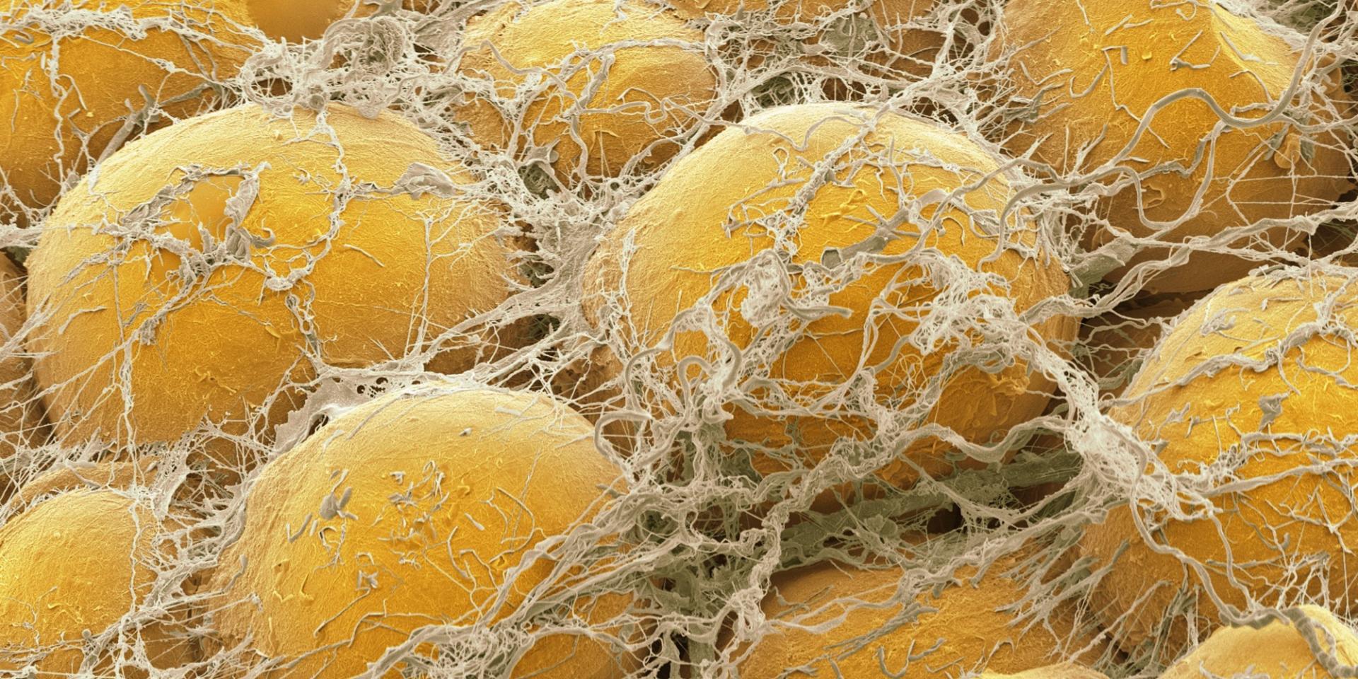
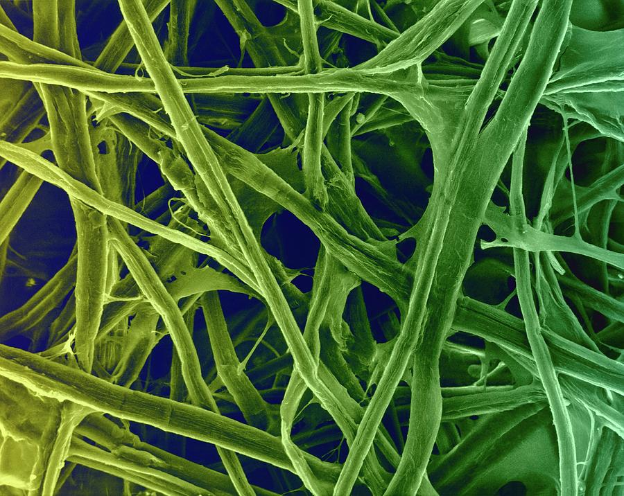
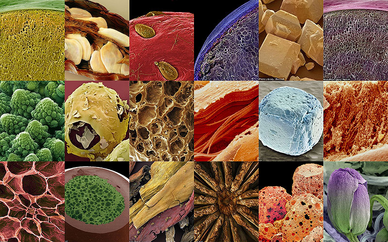
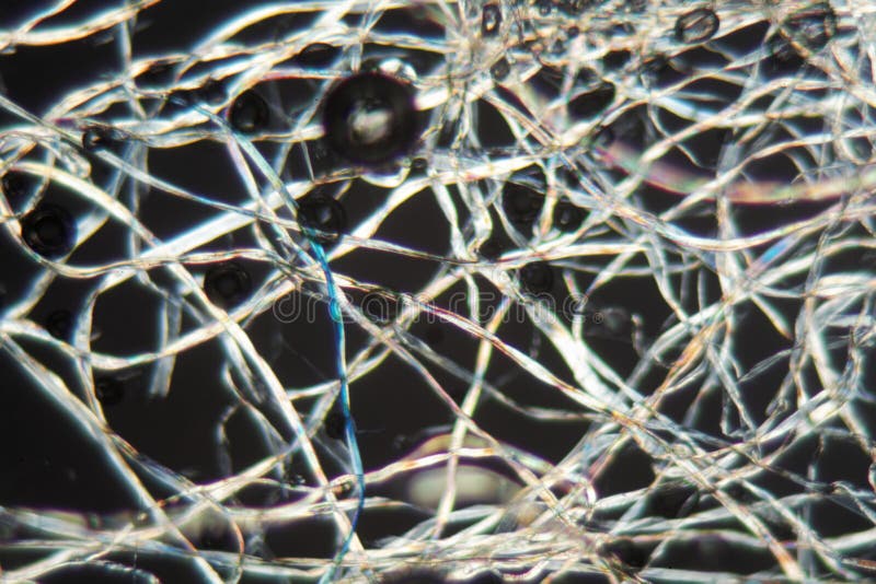
Как научить компьютер открывать новые материалы - все самое интересное на ПостНаМакрофотографии еды от Caren Alpert - DRIVE217 снимков еды под микроскопом Microscopic photography, Extreme close up, CoffeePin by Daniel Ryu on Life Under The Lens ( Geon Woo (Daniel)) LensCotton Fibers Under the Microscope Stock Photo - Image of close, material: 84441Микроскопный клуб. Мир под микроскопом ВКонтактеChemical and Electrical Synapses Biology for Majors IIAdipose (Fat) Tissue: Types, Benefits, and DisordersLOOK: Award-Winning Microscopic Images Science images, Microscopic photography, Pin on micro, macro, close-upЧто посмотреть под микроскопом? (часть 2) Наши дети Paper lamp, Novelty lamp, LaDiscover the Fascinating World of Microscopic FoodКартинки РАСТИТЕЛЬНАЯ КЛЕТЧАТКА ПЕРЕВАРИВАЕМАЯ В КАЛЕDiagram of potato cell QuizletFile:Yellow adipose tissue in paraffin section - lipids washed out.jpg - WikipedDiscover the Fascinating World of Microscopic FoodSolanum tuberosum Cells (potato) Diagram QuizletSperm Production Site, Sem #13 Photograph by Science Photo Library - Fine Art AmUnder a Microscope Even Familiar Things Look Beautifully Weird Mind blowing imagМикрофотография поперечного сечения нервного пучка. - Мы увеличиваем вещи как ниFantastic Voyage Electron Micrograph For the Home Photography, Micro photographyUnder the Microscope: Can you guess what these 10 images are? - Relatively InterPin on БіологіяA scanning electron microscope looks closely at the skin of a strawberry. #RainbInfinity Imagined Microscopic photography, Mixed breed dogs, Amazing natureТкань под микроскопом Zygar.ru Дзенсосудистые пучки папируса (Cyperus papyrus) в 200x увеличении Nikon small world,Фотоконкурс 2012 Wellcome Image Awards Moth fly, Science images, Micro photograpUC San Diego Health Sciences Spinal nerve, Scanning electron micrograph, MicroscВолос собаки под микроскопом (70 фото)Fascia Things under a microscope, Microscope, Electron microscopeО мембранах The North Face Futurelight - Блог "Спорт-Марафон"Coccidia at 400%. Very common in this area. Medical laboratory, Medical laboratoТридевятая земля (Невская Ксения) / Стихи.руStunning Microscopic Views of Everyday ObjectsCross Section of Grass - Smiley Faces Микроскопическая фотография, Микроскопы, ИChoroid Plexus Secretory Cells, SEM' Photographic Print - Steve Gschmeissner ArtУрок 10: Вещества неорганические - 100urokov.ruSpringtail. This is the skin surface of a spring tail (Collembola) with some haiШведские ученые при помощи холода остановили рост опухолейPin on scienceМакрофотографии еды от Caren Alpert - DRIVE2Peering into the micro world Electron microscope images, Scanning electron microЧто нужно знать о росте волос на теле человека ? Уникальное фото-как выглядит ро


:max_bytes(150000):strip_icc()/GettyImages-168835209-5669e2903df78ce16147bd5e.jpg)