Клеточная стенка под микроскопом - найдено 59 фото
Найдено фото: 59
moss-Sphagnum fimbriatum - Ohio Moss and Lichen AssociationPlant Epidermis Cellulose Cell Walls #2 Photograph by Science Photo Library - Fi
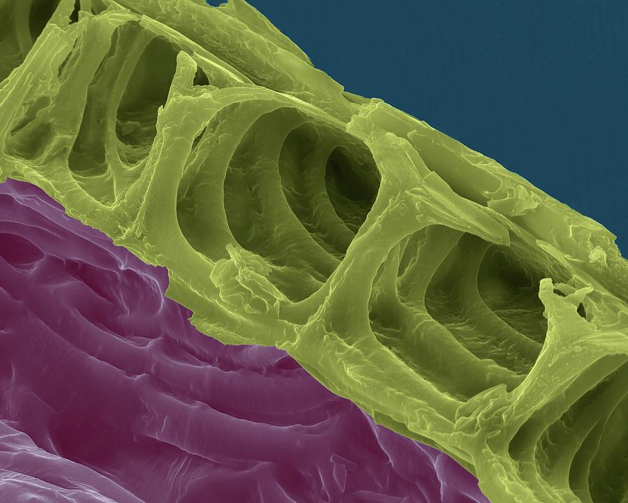
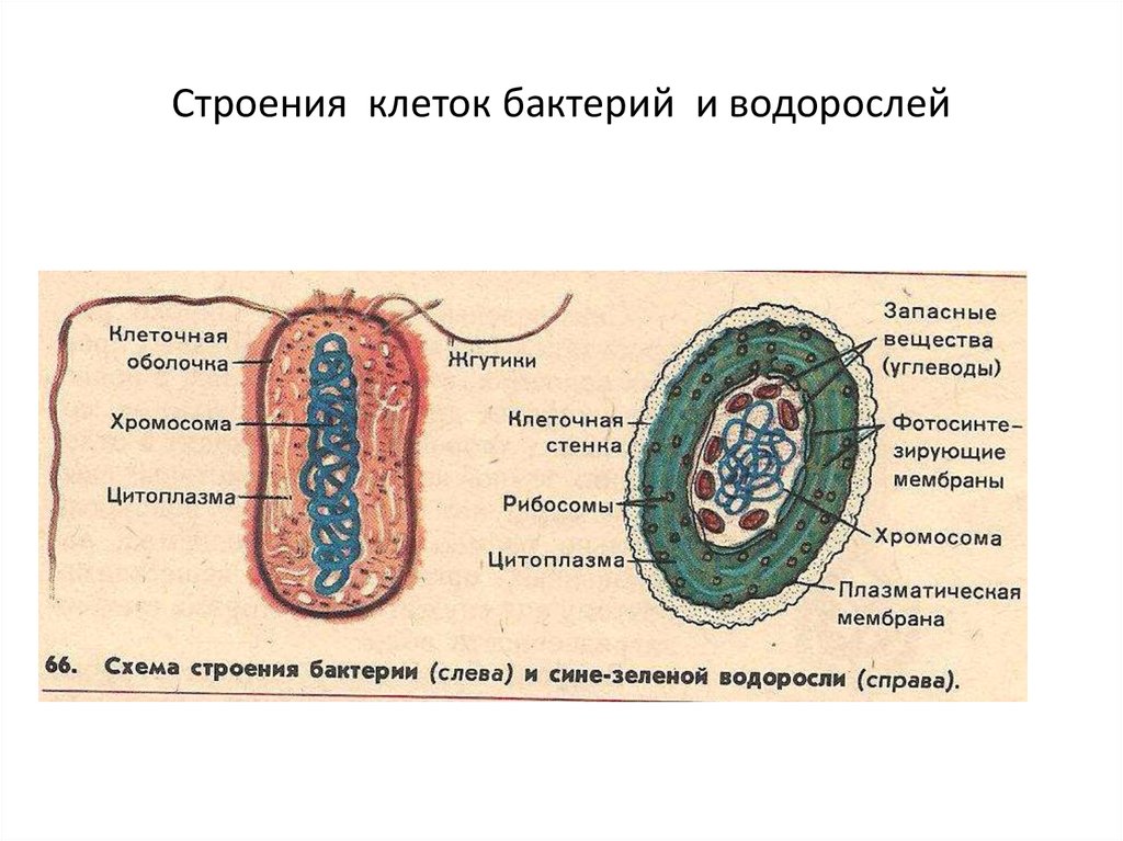
Biology careers - The University of SydneySchool of Engineering Vanderbilt UniversityРастительная клетка под световым микроскопом фото описаниеCell Wall Structure and Function Plant cell, Termites facts, Cell wallcell biology - Cyanobacteria: Gram negative or Gram positive? - Biology Stack ExXylem plant cells, SEM Plant cell, Microscopic photography, Frames for canvas pa
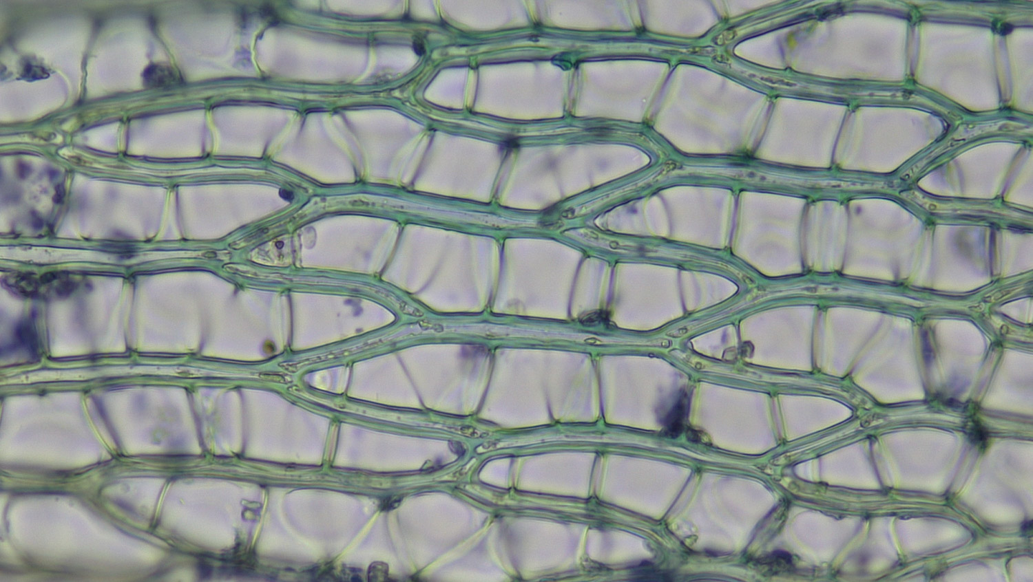
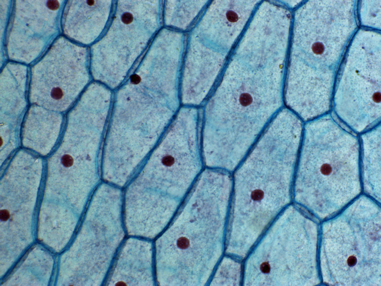
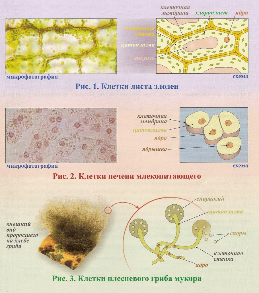
Bildergebnis für cell structure nature Microscopic photography, Microscopic cellКлетки защитной тканиMultimedia Gallery - Scanning electron microscope view of the water conducting tКартинки КЛЕТКИ РАСТЕНИЙ ПОД МИКРОСКОПОМThese award-winning microscope photos reveal a bizarre universe just out of reacElodea water plant under microscope. Cell walls and chloroplasts are clearly vis
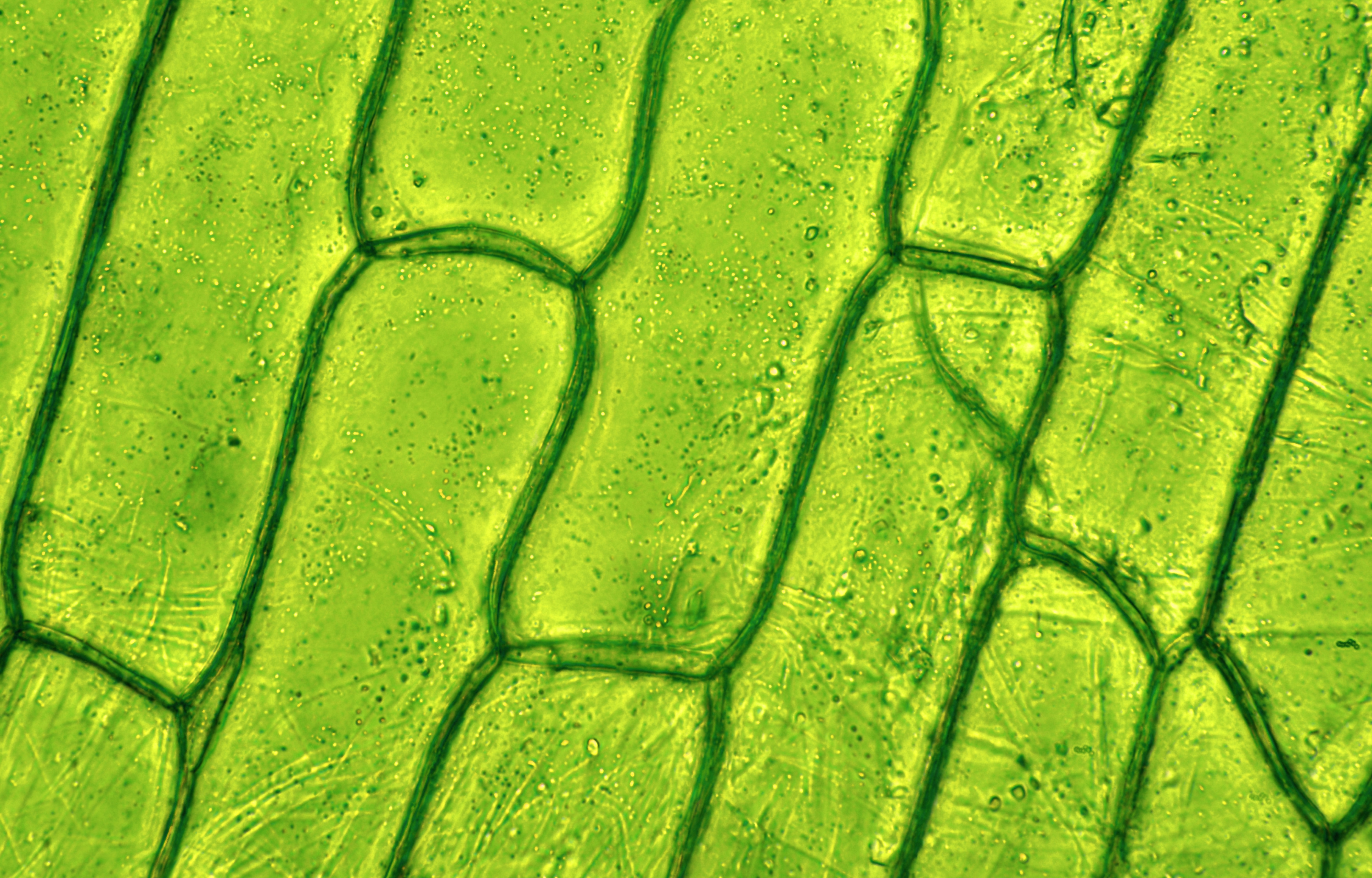
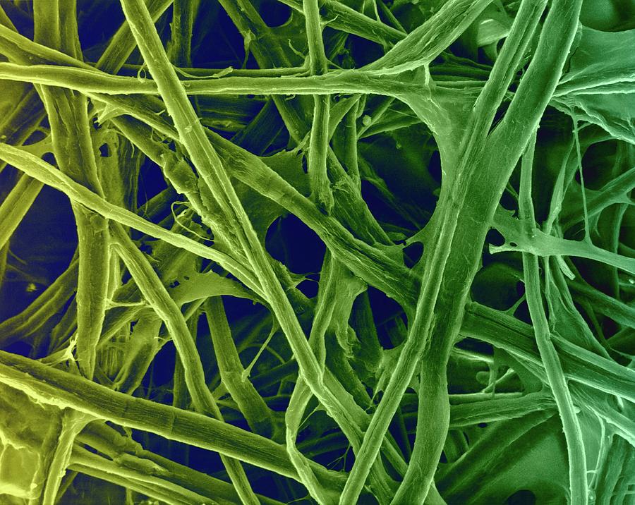
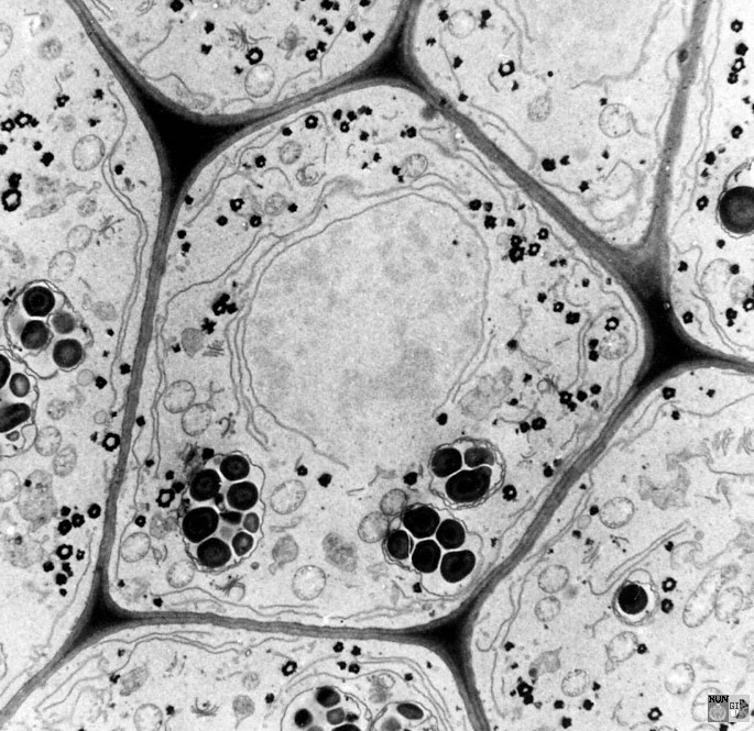
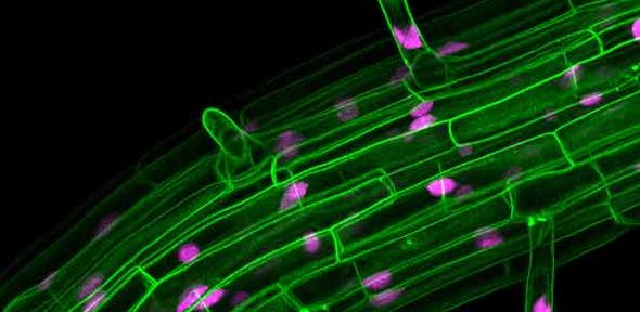
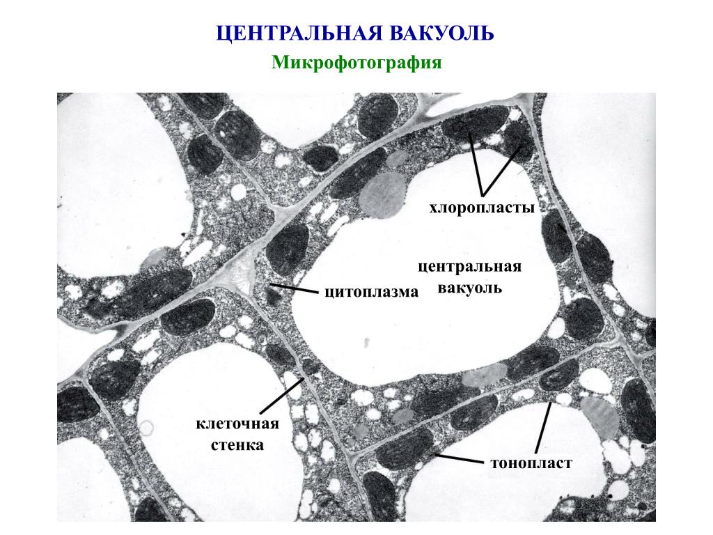
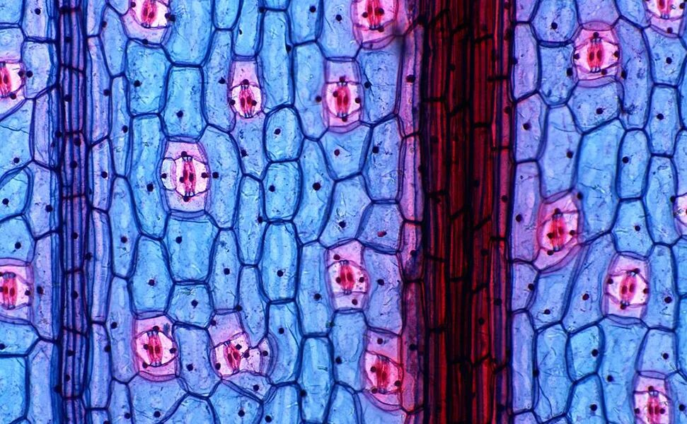
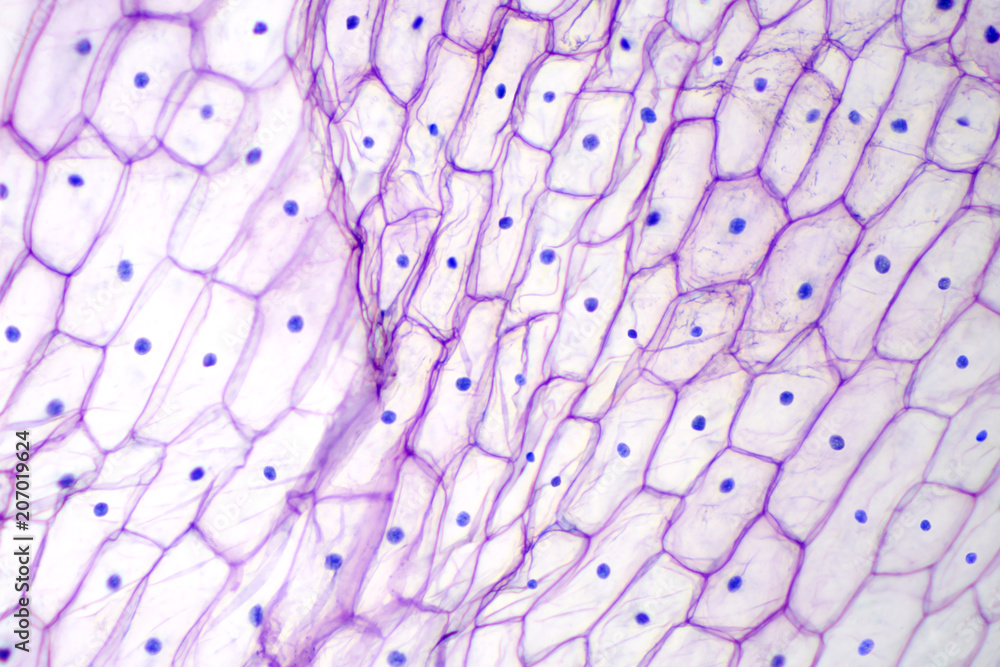
dictyosomesSearch in gallerycell Page 3 University of CambridgePin by Clay Stewart on microStudy Plant cell, Microscopic photography, Patterns xylem structures - Google Search Plants, Plant cell picture, Things under a micrEpidermis, Lcd, PixelBacteria lurking in blood could be culprit in countless diseases New ScientistEgeria_densa-saltwater Things under a microscope, Microscopic images, Science ceFile:Onion Cells.jpg - Wikipediapcell.gif (1045 × 1033) Photo, Aerial, City photoМикромир (фото) Electron microscope, Electron microscope images, Microscopic imaPPT - ВЕЗИКУЛЯРНАЯ СИСТЕМА КЛЕТКИ. PowerPoint Presentation - ID:6127435Оболочка растительной клеткиCloseup photo of Naples garlic Microscopic photography, Psychedelic plants, MicrКартинки КЛЕТОЧНЫЕ СТЕНКИ КЛЕТОКOnion epidermis with large cells under light microscope. Clear epidermal cells oМикрофотографии древесины Pellet associationOnion epidermis under light microscope. Purple colored, large epidermal cells ofDo Fungi Have Cell Walls? - Earthpedia Earth.com